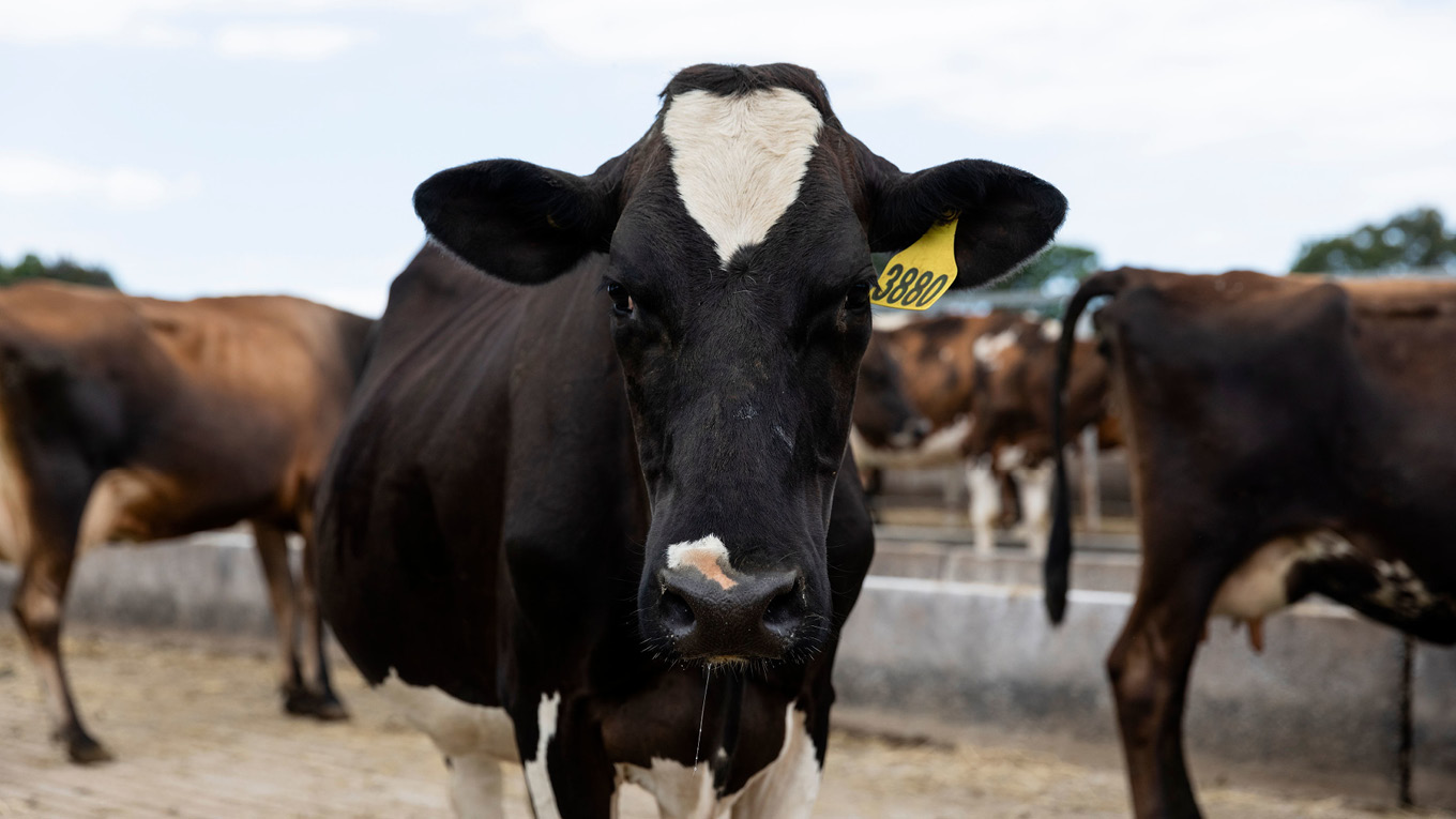
Sudden Death
Important:
Farmers should immediately report any unexplained sudden deaths of livestock to their veterinarian, state agriculture department or to the 24/7 Emergency Animal Disease Hotline on 1800 675 888.
If an animal that has died suddenly has blood coming from the nose, mouth or rectum, do not touch the carcass or remove any stock from the paddock.
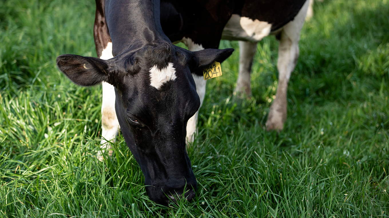
Causes of sudden death
Understand some of the more common causes of sudden death (with or without other signs) in dairy cattle.
Feed-related:
- Nitrate poisoning
- Ruminal acidosis
- Acute bovine liver disease (ABLD)
- Annual ryegrass toxicity
- Grass tetany
- Bloat.
Bacterial infections:
- Salmonella
- Anthrax
- Clostridial disease.
Other non-infectious causes:
- Polioencephalomalacia (PEM)
- Lead poisoning.
Rare causes of sudden death which are also discussed on this page include:
- Lightning strike
- Snakebite
Nitrate poisoning
What to look for
- Usually many animals affected at once
- Sudden death
- No sign of struggle
- Purple to muddy brown discolouration of tissue inside vulva.
Animals that are still alive may show
- Difficulty breathing and open mouth
- Staggering and muscle tremors
- Drooling
- Kicking at the belly, bellowing, lying down and diarrhoea
- Constant dribbling of urine
- Abortion.
Cause
Nitrate poisoning occurs when cattle graze plants that are taking up nitrogen faster than it can be converted to protein.
When consumed, high levels of nitrate undergo chemical reactions within the animal’s body which stop red blood cells from being able to transport oxygen.
Plants that have been associated with nitrate poisoning include ryegrass; cereals, e.g. oats, barley, maize, wheat; sorghum; millet; kikuyu; brassicas and some weeds, e.g. capeweed.
Younger plants are higher risk than mature plants. The concentration of nitrates is also higher in the stem or lower part of the plant than the leaf.
Animals likely to be affected
All dairy cattle consuming high-risk feeds may be affected. Hungry and stressed stock are more likely to be affected.
Confirming the diagnosis
Purple to muddy-brown tissues inside the vulva are diagnostic.
Fluid from the eye can also be tested for nitrates and nitrites in animals that have been dead up to 24 hours.
Testing of the feed source may also be useful to establish a diagnosis.
Treatment
Suspected nitrate poisoning is a veterinary emergency.
Remove all surviving animals off the affected pasture.
Vets may administer a product called methylene blue into the vein of any affected surviving animals. As methylene blue is not approved by the APVMA, written advice should be obtained about the treatment and withholding periods.
Risk factors
- Overcast weather
- Stressed plants (e.g. water stress, insect damage, frost, herbicides)
- Recent application of urea or other nitrogen containing fertilisers
- Grazing low to the ground
- Young, rapidly growing plants.
Prevention
- Regularly test high risk feed for nitrate levels via a vet or feed lab.
- Avoid grazing high risk feed during overcast or cold weather.
- Do not put hungry stock onto high risk feed, e.g. pre-feed with hay or silage.
- Check animals 1–2 hours after putting onto high risk feeds.
- Don’t graze high risk feed too close to the ground.
- Introduce high risk feeds slowly over time.
Cutting for silage or allowing plants to mature and set seed may reduce nitrate content in high risk feeds.
Ruminal acidosis
See ruminal acidosis section under feedbase.
Acute Bovine Liver Disease (ABLD)
Acute Bovine Liver Disease (ABLD) is an emerging disease of dairy cattle. Cases have now occurred in south-west Victoria, the Upper Murray, Tasmania, New South Wales and Western Australia.
What to look for
- Sudden death
- Photosensitisation such as sunburn and skin irritation
- Agitation and seeking shade
- Drop in milk production
- Depression and reduced appetite.
Signs are usually seen within hours of introducing cattle to high-risk paddocks.
Cause
The cause of ABLD is unknown. It usually occurs following a period of warm, moist weather when cattle are introduced to paddocks with high levels of dry feed and/or containing the annual grass 'rough dog’s tail'. Most cases occur in autumn/winter – April to July.
Animals likely to be affected
All cattle are susceptible to ABLD. Interestingly, horses and sheep do not appear to be affected.
Confirming the diagnosis
ABLD can be diagnosed by a veterinarian via blood tests or liver samples from live animals or via liver samples collected post-mortem.
Treatment
Move surviving cattle from suspect pastures immediately.
There is no known cure for ABLD. Treatment of affected surviving animals should be aimed at reducing pain and irritation associated with photosensitisation.
Risk factors
- A period of warm, moist weather or recent rain
- Introduction of cattle to pasture with high levels of standing and/or fallen dry feed
- The presence of the annual grass 'rough dog's tail' (Cynosurus echinatus).
Prevention
Wherever possible, avoid pastures with a previous history of causing ABLD particularly during high risk weather.
It is thought paddocks may remain toxic for up to two months following an ABLD event. Closely monitor cattle in at risk paddocks.
On some farms, it may be possible to pre-graze high-risk paddocks with sheep to reduce dry standing material.
Annual ryegrass toxicity
What to look for
- Sudden death
- Staggering, high-stepping and head wobbling
- Collapse and inability to rise
- Symptoms worsen when cattle are disturbed or stressed.
Cause
Annual ryegrass toxicity is caused by a neurotoxin. The neurotoxin is produced by bacteria that grow on the annual ryegrass seed heads.
Animals likely to be affected
All ages of cattle can be affected. Death rates can be high.
Confirming the diagnosis
Cattle may be affected between four days and four weeks of exposure to toxic pasture.
Diagnosis is usually made based on symptoms and exposure to annual ryegrass.
There is no specific diagnostic test for the condition.
Yellow slime (ryegrass gall) and abnormal, deformed seed heads may be observed on annual ryegrass plants. Failure to observe these changes does not rule out annual ryegrass toxicity.
Treatment
Surviving cattle should be quietly removed from affected pasture immediately and given supplementary feed and shade.
Deaths may continue for up to a week after cattle have been removed from toxic pasture.
Risk factors
Ryegrass toxicity occurs mostly in Western and South Australia between October and January when plants are maturing and forming seed heads.
Affected cattle are grazing annual ryegrass (Lolium rigidum) infected with the specific pasture bacteria and parasite.
Prevention
Affected pastures may remain toxic for a long period of time.
Hay made from infected pasture will also be toxic and should not be fed to stock.
Annual ryegrass toxicity may be prevented by:
- Using annual ryegrass species with increased resistance to annual ryegrass toxicity.
- Controlling unwanted annual ryegrass with post-emergent herbicides.
- Topping, hard grazing or mowing prior to seed head formation to prevent development of ryegrass gall.
- Spray topping to reduce annual ryegrass seed set and ryegrass population the following season.
- Burning affected pasture to destroy toxic material and seeds.
Grass tetany
See grass tetany section under down cows.
Bloat
See bloat section under diseases of the digestive system.
Salmonella
See salmonella section under diseases of the digestive system.
Anthrax
Important:
If an animal that has died suddenly has blood coming from the nose, mouth or rectum, call a vet, the local agriculture department or the Emergency Animal Disease Hotline on 1800 675 888. Do not touch the carcass or remove any stock from the paddock.
What to look for
- One or two animals and occasionally larger numbers are found dead
- Bleeding from the nose, mouth and rectum
- In the very early stages, animals have a high temperature, muscle tremors, difficulty breathing and/or convulsions.
Cause
A bacteria called Bacillus anthracis. The bacteria forms spores that allow it to survive in soil for decades. Animals become infected by eating or breathing in the bacterial spores.
Animals likely to be affected
Ruminants (cattle, sheep and goats) and horses are susceptible to anthrax. Anthrax is a rare disease in dairy cattle in Australia. Most cases have occurred in northern Victoria and inland NSW in a region known as the ‘anthrax belt’. Occasional cases have occurred in other areas of Victoria, NSW, Queensland and Western Australia.
Confirming the diagnosis
A diagnosis of anthrax is confirmed by laboratory examination of a blood sample that has been carefully collected from the carcass.
Treatment
Most animals with anthrax are found dead and severely ill animals are unlikely to recover. However, treatment is still recommended as it may reduce the number of infective spores released into the environment. Veterinarians may recommend that all animals with a high temperature greater than 39.5°C in affected herds should be treated with a course of antibiotics for at least 5 days.
Risk factors
Cases of anthrax often occur in warmer months and after heavy rain following hot dry weather.
Prevention
If anthrax has been confirmed, the affected property is quarantined to prevent further cases and potentially exposed stock are vaccinated.
Dead animals are safely disposed of, usually by burning, and contaminated sites are disinfected.
The quarantine is not released until at least 20 days have elapsed since the last anthrax case and at least 20 days have passed since the last round of vaccinations on the property, whichever is later.
Occasionally, larger-scale outbreaks occur, such as those in 1997 and 2007 in northern Victoria. Vaccination across a wider area is usually required to control larger outbreaks.
Farmers in anthrax-prone areas should contact their state agriculture department if they wish to undertake voluntary preventive vaccination against anthrax.
Anthrax in people
People can become infected with anthrax via contamination of cuts and scrapes when handling contaminated material, e.g. carcass of an animal that has died from anthrax. Carcasses of dead animals should always be handled with care using protective clothing and good personal hygiene.
Anthrax infection in people usually only results in skin infections in the form of pimples and boils and treatment with antibiotics is usually successful. Rarely, infection may occur from breathing in the anthrax bacteria. This can result in very severe pneumonia that can be fatal. This form of anthrax was once called wool sorter disease because it occurred in people who handled wool from dead sheep.
It is important to seek immediate medical attention if a skin infection develops or a person becomes unwell after potential exposure to anthrax.
Clostridial disease
There are several clostridial diseases that can affect dairy cattle. These include:
- Blackleg
- Malignant oedema
- Black disease
- Pulpy kidney
- Tetanus
- Botulism (discussed separately in conditions of the nervous system).
What to look for
- Sudden death
- Rapid decomposition and/or bloating of the carcass
- Very unwell animals with a high temperature
- Tetanus: Stiff gait, seizures and/or convulsions
- Blackleg: Lameness and swelling
- Pulpy kidney: Abdominal pain
- High temperature greater than 39.5°C.
Cause
Clostridial diseases are caused by infection with anaerobic bacteria of the Clostridium species. The bacteria are widespread in the soil and can survive in the environment for a long time. Some Clostridium bacteria are also found in the digestive system of healthy animals and infection occurs when conditions are favourablem for example changes in diet, lush feed.
Animals likely to be affected
Clostridial diseases often occur in well-fed, well-conditioned or growing animals.
Confirming the diagnosis
Diagnosis requires post-mortem examination by a veterinarian.
Treatment
In very early cases, treatment may be attempted with appropriate antibiotics but is often unsuccessful.
Risk factors
- Non-vaccinated animals
- Wounds including those associated with husbandry procedures e.g. disbudding, castration
- Muscle bruising
- Liver fluke infection
- Lush pastures or high concentrate diets
- Recent calving.
Prevention
Clostridial diseases except botulism are prevented by vaccination with either 5–in–1 or 7–in–1.
Vaccines should always be used according to the label directions.
Most vaccines recommend an initial course of two vaccines 4–6 weeks apart from 6 weeks of age followed by an annual booster.
Ideally, vaccines should not be given when animals are exposed to other sources of stress, for example at disbudding or weaning, as this may reduce the efficacy.
Vaccines should be kept refrigerated or put in an esky with an ice brick and out of direct sunlight during transport and when not in use. Do not freeze vaccines.
It is important not to forget carryover cows when booster vaccines are typically given at dry off.
Botulism is not covered in 5–in–1 or 7–in–1 but can be prevented by a different vaccine.
Polioencephalomalacia (PEM)
What to look for
- Sudden death
- Depression, blindness and/or staggering
- Pressing head against solid objects
- Abnormal chewing behaviour or teeth grinding, drooling
- In severe cases, animals will go down and exhibit rigid muscles and convulsions before dying.
Cause
Polioencephalomalacia (PEM) is caused by a thiamine deficiency or excessive sulphur intake.
Animals likely to be affected
PEM usually affects well-grown calves between 6 and 12 months of age. It can occasionally affect older cattle following severe ruminal acidosis.
Confirming the diagnosis
Confirming the diagnosis of PEM in live animals is difficult and largely relies upon response to treatment.
Often the history of the animal is suggestive.
Post-mortem examination of the brain from an animal that has died or been euthanased can confirm the diagnosis.
Treatment
Treatment involves large doses of thiamine being given into the vein by a veterinarian followed by regular intramuscular thiamine injections for a couple of days. However, even with treatment, the likelihood of recovery can be difficult to predict.
Risk factors
- High concentrate, low fibre diets
- Sudden changes in diet
- Drinking water with high sulphur content
- Exposure to bracken fern.
Prevention
PEM outbreaks are difficult to predict and therefore prevention is challenging.
Always introduce new concentrates and/or new rations gradually to allow for rumen adaptation.
If the cause of PEM on an affected farm can be identified, it should be addressed.
Seek an alternative water source if sulphur levels exceed 500 ppm.
Lead poisoning
What to look for
- Sudden death or death within 24 hours of developing symptoms
- Staggering and blindness
- Muscle twitching, extreme sensitivity to touch
- Frothing at the mouth
- Abnormal chewing behaviour or teeth grinding
- Mild cases may exhibit dullness, reduced appetite, abnormal movement, diarrhoea.
Cause
Lead poisoning occurs when animals have access to paint containing lead, e.g. on the walls of old buildings, lead-acid batteries or waste motor oil.
Animals likely to be affected
Most cases of lead poisoning in dairy cattle occur in calves less than six months of age due to their natural curiosity and tendency to taste everything.
Confirming the diagnosis
Lead poisoning is often diagnosed by the presence of symptoms in combination with exposure to lead.
A blood test is available to confirm the diagnosis.
Post-mortem diagnosis of lead poisoning is very difficult.
Treatment
Animals may recover from lead poisoning if they receive veterinary treatment at an early stage.
Young animals are less likely to respond to treatment.
Risk factors
Exposure to buildings painted with paint containing lead as well as lead-containing materials such as lead-acid batteries, lead flashing or waste motor oil.
Prevention
Avoid exposure, particularly of young animals, to lead-containing materials.
Lightning strike
What to look for
- Sudden death in a single animal or group of animals close to a fence, tree(s) or water
There may also be:
- Signs of struggle
- Bleeding from the nose, mouth and eyes
- Burns or singing of the hair, nose or feet.
Cause
Lightning strike causes heart and/or respiratory failure by electrocution.
Confirming the diagnosis
Animals that have bleeding from the nose, mouth, eyes or other body cavities should not be touched or moved and a veterinarian contacted immediately to exclude anthrax.
Often there is a history of thunderstorms with carcasses found under trees, near fences or water.
However, post-mortem examination by a vet can help to confirm the cause.
Many insurance policies cover lightning strike and examination by a veterinarian is required to process claims.
Treatment
Animals suffering from lightning strike are almost always found dead.
Animals that were not killed may be showing blindness, staggering or paralysis.
These animals may recover with supportive care.
Risk factors
- Thunderstorms
- Risk appears higher for tall or hardwood trees with sprawling root systems, e.g. oaks, eucalypts.
Snakebite
Contrary to popular belief, snakebite in cattle in Australia is actually extremely rare due to the large size of cattle relative to the dose of toxin required to cause death.
What to look for
Signs of snake bite in cattle may include
- Sudden death
- Cows may be paralysed and go down
- Painful swelling at bite site
- Behavioural excitement or agitation
- Dilated pupils, drooling and a floppy tongue.
Confirming the diagnosis
Occasionally snakebite will be witnessed, e.g. a snake in the feed trough of the dairy, or the snake will be still attached to the animal.
Snakebite can be confirmed by blood tests conducted by a veterinarian.
Post-mortem may occasionally support the diagnosis in an animal found dead.
Treatment
Snakebite can be treated with antivenom. However, antivenom is very expensive so its use may be limited to high-value animals.
Identification of the species of snake is not required as most antivenom in veterinary clinics is polyvalent, meaning it works against the venom of most common snake species.